
Adult stem cells are present in almost every tissue in our body, and they are responsible for maintaining the tissue homeostasis throughout life. The focus of research in this laboratory is to understand the basic biology of adult stem cells in the human eye, elucidate changes with ageing/diseased conditions and develop better stem cell based therapies. Studies are being carried out on (i) trabecular meshwork stem cells in relation to primary open angle glaucoma, (ii) lens epithelial stem cells and age-related cataract, and (iii) retinal pigment epithelial stem cells and age-related macular degeneration.
Previous reports from this laboratory have confirmed the location of stem cells for trabecular meshwork in the anterior non-filtering region, lens epithelium in the central zone, and retinal pigment epithelium in the peripheral region.
- Molecular regulation of stem cells: Studies are now being carried out to understand the molecular regulation of these adult stem cells, including transcription factors, microRNAs and associated signalling pathways. Identification of these intrinsic factors will enable the development of better methods for expanding the stem cells in vitro as well as to activate them in vivo.
- Changes in adult ocular stem cells with ageing/diseased condition: Quantification of the stem cells revealed a significant reduction in trabecular meshwork stem cells in glaucomatous condition, functional loss of lens epithelial stem cells in age-related cataract, and retinal pigment epithelial stem cells with ageing.
- Towards stem cell-based therapy for glaucoma: Recently, exosomes derived from the trabecular meshwork stem cells were demonstrated to have enhanced wound healing and anti-oxidant potential in vitro, indicating the possibility of developing a cell-free therapy for glaucoma. Further studies are being initiated to evaluate the efficacy of these nanovesicles in TM regeneration using animal models for primary open angle glaucoma.
In addition, projects of clinical significance carried out in this department are:
- Transplantation of autologous limbal/buccal mucosal epithelial stem cells for corneal surface reconstruction in patients with limbal stem cell deficiency
- Evaluation of the efficacy of the corneal storage medium – Cornisol
- Effect of VEGF level in Tenon’s fibroblasts on the outcome of glaucoma surgery
- Evaluation of surface free energy of the glaucoma drainage implant (AADI)
- Mechanism of understanding posterior capsular opacification (PCO)
Ongoing Projects
- Molecular regulators associated with the maintenance of human trabecular meshwork stem cells in relation to their reduction in ageing and glaucoma – SERB project (2021-2024)
- Molecular characterisation of human retinal pigment epithelial stem cells and their role in age related macular degeneration – ICMR project (2022-2025)
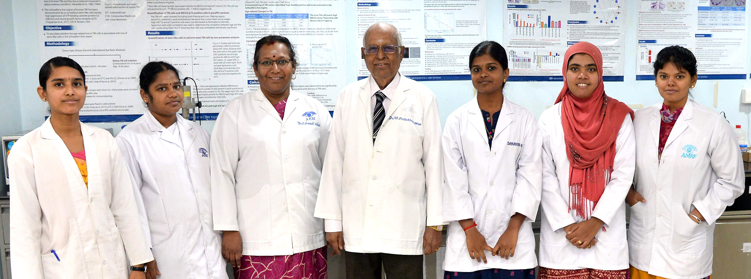
Faculty
Ramalingaswami Fellow
Research Scholars
- Ms. P. Saranya
- Mrs. A. Waseema
- Mrs. R. Iswarya
- Ms. S. Sneha Nair
- Ms. T. Keerthana
- Ms. P. Thushmitha
- Ms. R. Kanthimathi
- Ms. Gopika S. Kumar
Lab Assistants:
- Mrs. P. Vigneswari
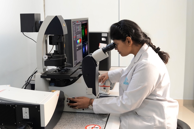
Confocal Laser Scanning Microscope

Model: TCS SP8
Manufacturer: Leica, Wetzlar, Germany
Leica TCS SP8 confocal laser scanning microscope is an inverted microscope designed for optical imaging with optimal photon efficiency and high speed; facilitating optical sectioning. This microscope is equipped with 4 laser ports namely UV/405, laser blue 488nm, laser green 552nm and laser red 638nm.
Laser Assisted Microdissection System
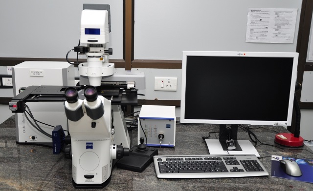
Model: PALM Microbeam IV
Manufacturer: Carl Zeiss, GmbH
This system enables laser-assisted isolation of individual/group of cells from fixed sections/live cultures that can be used further for genomics/proteomics