
The Proteomics department focuses on ocular diseases of national importance, such as fungal keratitis, keratoconus, and pterygium, as well as globally relevant conditions like diabetic retinopathy and glaucoma. We aim to understand disease pathogenesis and develop diagnostic, prognostic, and therapeutic methods to improve patient care. We are working on non-invasive therapies to enhance current treatments. Our research on diabetic retinopathy discovered and validated a panel of circulatory biomarkers. We collaborate internationally with institutions like the University of Edinburgh (fungal keratitis), Moorefields Hospital (diabetic retinopathy), and the University of Liverpool (keratoconus).
Fungal Keratitis
Fungal keratitis accounts for over 50% of infectious keratitis cases, with Fusarium and Aspergillus flavus being the predominant pathogenic species. A multi-OMICS approach has been used to study themycotic keratitis pathogenesis in both human host and fungal pathogens. Genomics, transcriptomics, and proteomics of the fungi have provided insights into species differences, while host proteomics of tear and cornea reveal key pathways activated during infection. We have identified tear proteins as markers of infection severity. We are now evaluating the extracellular vesicle (EV) protein profiles from tear samples to develop EV-based biomarkers for predicting fungal ulcer prognosis. Additionally, we’re studying fungal EVs contributing to disease pathogenesis.
Diabetic Retinopathy
Diabetic retinopathy (DR) is a leading cause of vision loss globally, especially in working-age individuals. In India, DR affects 12.5% of the diabetic population, with 4% experiencing vision-threatening DR (VTDR). Early-stage DR is often asymptomatic, only becoming evident at advanced stages like proliferative DR or diabetic macular edema. Our research aims to identify protein biomarkers for DR diagnosis and prognosis. We have analysed serum and vitreous proteomes to identify stage-specific proteins, focusing on circulating microparticles and microRNAs. In an Indo-UK collaboration, we validated serum markers in Indian and UK populations to detect high-risk patients and predict DR progression. We’re also studying EVs from plasma and vitreous fluid as potential diagnostic markers.
Glaucoma
Glaucoma is the leading cause of blindness worldwide. We are analysing the mutations responsible for the occurrence of early and late-onset POAG (Primary Open Angle Glaucoma) in members of large families. The effects of these mutations at the cellular level will be analysed in-vitro. Proteome analysis of aqueous humour from POAG patients enabled the identification of protein changes specific to POAG. These proteins will be validated in a larger patient cohort to develop a panel for diagnostic or prognostic use.
Keratoconus
Keratoconus is a corneal disorder prevalent in India, causing thinning, weakening, and severe vision defects. Factors like genetics, intraocular pressure, and collagen weakening contribute to its progression. We have developed a novel eye-drop-based chemical cross-linker to treat keratoconus, aiming to simplify treatment by avoiding the surgical removal of the epithelium and pain associated with conventional UV-Riboflavin cross-linking. In collaboration with AMRF, the University of Liverpool, and Aurolab, the cross-linker has shown low cytotoxicity in human corneas and rabbits, significantly increasing corneal stiffness without altering morphology. It inhibits matrix metalloproteases, showing promise for further trials and formulations, with Aurolab handling the final clinical development.
Pterygium
Pterygium is a common conjunctival eye disease, primarily affecting outdoor workers from low socioeconomic backgrounds, impairing vision and quality of life. UV exposure is a major risk factor, but the exact cause is unclear. With a prevalence of 12% and no pharmaceutical treatments, surgical removal is the only option. Our research integrates proteomics and transcriptomics of conjunctival samples from pterygium surgeries to uncover key molecular pathways involved in disease progression. We aim to develop small-molecule inhibitors to halt progression. We are also exploring genome-wide methylation changes to understand the role of epigenetic alterations in pterygium pathogenesis.
Ongoing Projects
- Deciphering predictive and preventative methods in the progression of pterygium using multiomics approaches. (SERB-SRG, Dr. Daipayan Banerjee)
- Role of Extracellular Vesicles in Diabetic Retinopathy: A comparative proteomic analysis of plasma and vitreous humour derived small Extracellular Vesicles (SEVs) from Proliferative Diabetic Retinopathy (PDR) patients (SunPharma)
- Understanding the mechanism of action of a novel chemical cross-linker designed to treat keratoconus (ICMR, Dr. O.G. Ramprasad)
Faculty
- Prof. K. Dharmalingam, Director – Research
- Dr. O.G. Ram Prasad, Scientist
- Dr. Daipayan Banerjee, Scientist 1
Research Associate- II
- Dr. T.M. Viswanathan
Project Associate – II
- Karthik Alagarsamy
Research Scholars
- Aadhithiya. T. Gr
- Mathan Loganathan
- Hariharan Gnanam
Lab Assistants
- T. Parameshwari
- M. Sujitha
- M. Manikandan
- M. Dhanalakshmi
Mass spectrometry facility
The core proteomics facility has two mass spectrometers.
- nanoLC-Orbitrap velos pro mass spectrometer: This is a high-resolution mass spectrometer that enables the in-depth identification and quantitative comparison of proteins in a complex proteome such as tears, serum, corneal tissue, fungal cells, etc.
- uHPLC/nanoLC-Triple stage Quadrupole mass spectrometer: This mass spectrometer is used for a high-throughput, reproducible, and targeted quantitation of proteins in complex proteome
Exosome Innovation Centre
We have established a state-of-the-art facility to facilitate exosome research in ocular and other related areas. This facility is a part of the translational initiative for the basic research programmes at AMRF. We are expanding the scope of this exosome research facility to be available for any group working in the exciting yet challenging area of exosome research. Our facility includes the infrastructure for carrying out the end-to-end workflow, right from isolation to confirmation to characterisation of exosomes.
This facility encompasses the major infrastructure essential for the entire workflow.
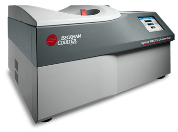
Beckman-Coulter tabletop ultracentrifuge for exosome isolation
The Optima™ MAX-XP is a tabletop ultracentrifuge that delivers fast, efficient separations from samples as small as 175 μL up to 32.4 ml and at speeds of up to 150,000 RPM and more than 1,000,000 x g. This facility is equipped with three different rotor types – fixed-angle MLA-130 rotor (for sample volumes less than or equal to 1 ml), fixed angle MLA-50 rotor (for larger sample volumes >7 ml), and swing bucket MLS-50 rotor.
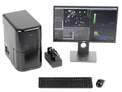
Malvern NanoSight NS300 for nanoparticle tracking analysis
Nanoparticle Tracking Analysis (NTA) utilises the properties of both light scattering and Brownian motion in order to obtain the nanoparticle size distribution of samples in liquid suspension.
The NanoSight NS300 instrument is a laser-based, light scattering system for specific and general nanoparticle characterisation. With the NS300, it is possible to analyse the presence, size distribution, concentration, and fluorescence of extracellular vesicles from 10 nm to 2000 nm.
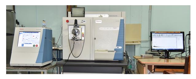
nanoLC-Orbitrap Velos Pro Mass Spectrometer for proteome profiling
The Mass Spectrometry facility includes a fully integrated nano-LC system that works up to 1000 bar (15000 psi.) and a narrow column ID to increase analyte and improve detection sensitivity. This front end seamlessly integrates with the Orbitrap Velos ProTM mass spectrometer, which combines Orbitrap ™ mass analyser and Velos Pro ion trap technology to deliver high resolution, speed, sensitivity, and flexibility.
Other facilities
Additionally, a cell culture facility, a clean room for fungal culture, a clean lab for RNA work and qPCR are available.
Services provided
Users can opt for services for the entire workflow or any one or more of the services listed below.
1. Isolation of exosomes by the ultracentrifugation method
Ultracentrifugation is the gold standard method for isolating exosomes. We have already optimised UC based isolation of exosomes from plasma, tears, vitreous humour, cell culture spent medium, and fungal culture. Hence, we can help isolate exosomes from various sources such as,
a. Any biological samples such as serum, plasma, tears, urine, milk, etc.
b. Culture supernatant from mammalian cells, bacterial culture, and fungal culture
2. Confirmation of exosomes by Nanoparticle Tracking Analysis (NTA)
It is important to confirm that the isolation protocol enriched the exosomes from the starting material and NTA is routinely used for this purpose. NTA will provide valuable information that is not only required for confirmation but also for downstream functional assays. As a part of the exosomes workflow, after isolation, we perform NTA analysis to obtain information on:
- Size distribution
- Number of exosomes per ml of the starting material
- Enrichment of exosomes (Count vesicles before and after the UC step)
3. Characterisation of protein cargo in the exosomes
Exosomes harbour various cargo – proteins, RNA, miRNA, and lipids. The protein cargo of the exosomes will be quantified, identified, and characterised as follows:
- Concentration of proteins in the exosomes – spectrophotometric quantitation using the BCA assay (will be performed only if sufficient yield of exosomes are achieved)
- Exosome protein profiling
- Depending on the concentration and number of exosomes available, proteome profiling will be carried out either using in-solution or shotgun in-gel tryptic digestion.
- Extraction of peptides and desalting using C18 columns
- Analysis of tryptic peptides in nanoLC-mass spectrometer
- Processing of MS raw data to list of proteins
4. Additional work elements
-
- Protein profiling from tissues, body fluids, and cells
- Extended analysis of the proteome data
- Consultation on experiment design for exosome research
Further details will be worked out upon discussion with the user.