
Glaucoma is the second leading cause of blindness, affecting 70 million people worldwide, and there is always a constant search for the discovery of newer/better drugs for glaucoma. Our lab has developed a Perfusion cultured human anterior segment (HOCAS) system to determine human trabecular meshwork outflow facility in response to investigational drugs. This ex vivo model system has been successfully exploited for various applications such as screening of drugs for glaucoma, creation of disease models [steroid-induced OHT model, saponin-induced cell loss model, and TGFβ2-induced glaucoma fibrosis model] and development of miRNA-based therapeutics for glaucoma. The other research areas include ocular pharmacokinetics, drug analysis by HPLC, pre-clinical in vitro /ex vivo model for eye diseases and retinal neuro-degeneration.
Development of Human Relaxin (RLX2) as a potential anti-fibrotic agent for glaucoma
Relaxin is a heterodimeric peptide hormone with known vaso-dilatory and anti-fibrotic activity. Our previous study suggested the involvement of relaxin in steroid sensitivity. However, the role of relaxin and its non-allelic gene variants in human aqueous outflow facilities and IOP regulation is not clearly understood. Considering the distribution of relaxin and its receptors in human aqueous outflow pathway tissue, it is hypothesised that relaxin 2 may have an active role in regulating outflow facility and IOP in the human eye. Currently, a TGFβ2-induced elevated IOP ex vivo model of glaucoma was established to demonstrate the IOP lowering property and anti-fibrotic properties of relaxin 2. We believe that relaxin 2, with proven IOP lowering and anti-fibrotic properties, will be more beneficial in the management of glaucoma.
Human Organotypic Retinal Explant Cultures (HORCs) for investigating neuroprotectants
Organotypic explant cultures allow controlled experimentation of retinal tissue and have the advantage over dissociated cultures by retaining regional morphology. Human Organotypic Retinal Cultures (HORCs) serve as a suitable model for investigating the RGCs after excite-toxicity. Currently, the neuro-protective property of GSK inhibitors and other molecules using this model is in progress.
Ongoing Projects
- Evaluation of IOP lowering property and Anti-fibrotic property of Relaxin on TGFβ2-induced Elevated IOP Ex vivo model of glaucoma using Human Organ Cultured Anterior Segment (HOCAS) – SERB-CRG grant
- Decoding the Unknown Genetic Etiology of Leber Congenital Amaurosis and Assessing the Complex Factor to Implement the Gene Therapy Trials in India- DHR grant
- ‘Relaxin’ the pressure in glaucoma – Fight for Sight PhD Studentship 2022 programme (UK)
- Role of vitamin D3, dopamine and serotonin levels in myopia development and progression – Aravind-Madurai
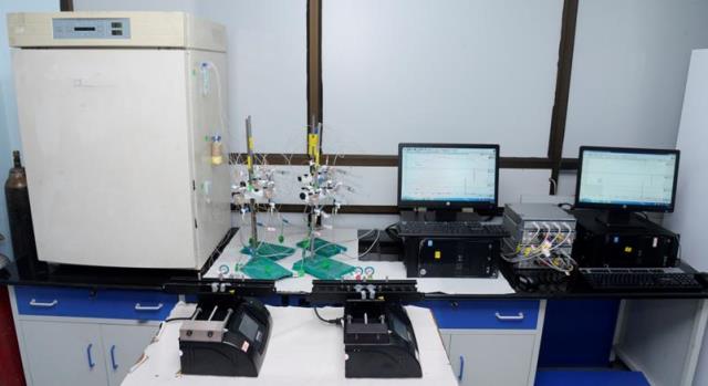
Human Organ Culture Anterior Segment (HOCAS) Facility for Trabecular Meshwork Outflow Studies
The human organ cultured anterior segment system (HOCAS) has been developed as an intermediate step between cell culture and whole animal or human studies for determining trabecular meshwork outflow facility responses to investigational drugs. This system allows direct experimentation of candidate drugs on human/bovine/pig and monkey eyes and thus provides direct and meaningful information about the candidate drugs for glaucoma. They are also useful for investigating the potential of gene therapy to target the trabecular outflow pathway.
HOCAS: Instrument Set-up
High Performance Liquid Chromatography (HPLC) for bio-analysis of Drugs
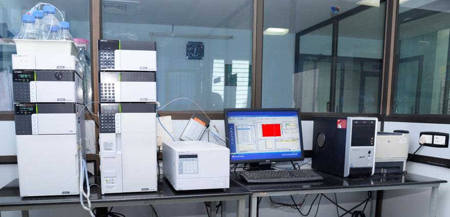
Equipment Setup
Shimadzu Prominence HPLC system with PDA detector (Shimadzu Corporation, Kyoto, Japan).
Tissue culture Facility
Our department maintains cell lines and primary cultures as follows:
- Tissue and cell culture: Maintenance of ARPE-19 cell lines in monolayer.
- Primary culture (e.g. Retinal Pigment epithelial cells and Trabecular Meshwork cells from human donor eyes obtained from Rotary International Eye Bank of Aravind- Madurai).
- Human organotypic Retinal Explant culture (HORCs) for screening neuroprotective agents for glaucoma
Services:
- Human Organ Culture Anterior Segment (HOCAS):
Hands-on training will be done on Dissection, HOCAS set up, Drug treatment protocol, and data analysis. This facility can be used for functional studies in the pathogenesis of glaucoma. - Pharmacokinetics of drugs and estimation of metabolites using HPLC
- Drug response and functional studies using primary culture and cell lines